 |
|
Pollen grain of a lily Click and wait - sequence will be loaded. |
CONFOCAL MICROSCOPY
The image obtained using an optical microscope unfortunately consists of three images which have merged into one:
1. The image of the focal plane.
2. The images of the planes above this plane which are out of focus.
3. The images of the planes below this plane which are out of focus.
Obviously the images out of focus blur the resulting image.
In 1957 Marvin Minsky suggested to use the confocal principle, and in 1967 the first commercial microscope using this principle was introduced onto the market.
The method is basicly simple, but the realisation is quite sophisticated: A bright light source is focussed on a very small point of the sample, then the intensity of the passing light (bright field microscopy) or of the reflected light (reflected light microscopy) is measured by an electronic detector. This kind of illumination reduces the above mentioned blurred images to an absolute minimum. In order to get an image the sample must be scanned first, then the resulting pixel values are reassembled. A very small aperture ("pinhole") close to the detector reduces the depth of field to a few hundred nanometres, so that an optical section of the sample is obtained.
The optical resolution of such microscopes is limited by the "Abbe´s formula", but now a factor of this formula, dealing with the contrast sensitivity of the detector and usually omitted, becomes important leading to a possible increase of limiting resolution of about 30%. Much more important, however, is the dramatic increase of clarity combined with a much better depth of field when many optical sections are merged using stacking programs. This has become possible because good laser sources and powerful computers are now available. For amateurs, however, such microscopes would normally be too expensive.
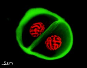
Today confocal microscopes are usually designed as fluorescence microscopes using reflected light illumination. The resulting quasi-3D images are converted to false colour images afterwards.
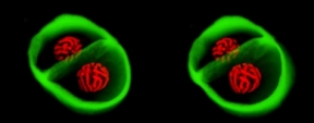
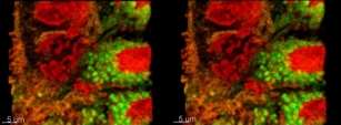
Mr.Wolfgang Posselt, a former member of our group, deals with the creation of such images professionally. Here you can see the image of the pollen grain of a lily.
|
|
Pollen grain of a lily Click and wait - sequence will be loaded. |
Extracting two images of the whole
sequence using the program VIRTUAL DUB makes it possible to assemble them to
stereo pairs. Click on the pair and look at the larger version from reading
distance cross-eyed - the right eye will then see the left image and the other
way roun. In the middle of the pair you can see two overlapping images which
will completely merge after some seconds resulting in a real stereo image. Our
brain will fix the orientation of the eyes then, so you can look at the 3D-image
without any stress. Having been successful once, this method will always work
effectively.
To get more information use the links iven below:
www.mh-hannover.de/lasermicroscopy.html
and