HE-staining
MASSON-GOLDNER-
staining
Sclera and conjunctiva
MASSON-GOLDNER-
staining
Very small fish such as guppies
are particularly suited for microscopic studies, since their hard parts are
still largely unossified and the objects can easily be cut with the microtome.
All the paraffin sections are self-made
(MINOT microtome, section thickness of 8 um), the pictures were made with the
combination of 10x5 (PK-projective), 25x5 (PK-projective) and 40x5 (PK-projective)
(objectives LEITZ achromates). Camera type: Kodak EasyShare C613, 6.1 megapixels.
The images shown here have been compressed, but because of the inherent fuzziness
of microphotographs of this kind the loss of quality is hardly susceptable.
For more information see Digital Photography
.
|
Caption | |
|
|
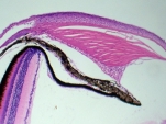
| Iris HE-staining |
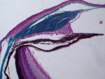
Iris MASSON-GOLDNER- staining |
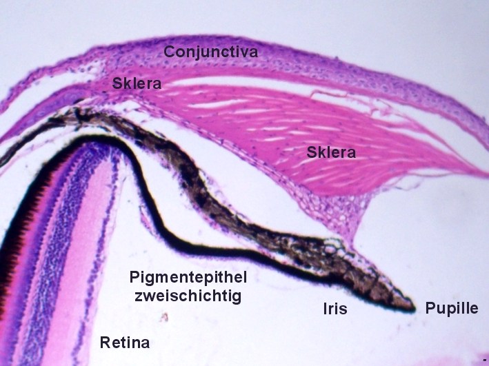
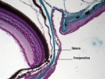
Sclera and conjunctiva |
The examination of the eyes
is always most interesting.The pictures above show the structure of the iris.
We can clearly see that the iris is made of three layers: Inside there are two
layers filled with pigment, outside superposed by another thin layer
of tissue, this is clearly visible on the left picture. The human iris has the
same construction: if the outer layer is relatively thick, the eyes appear grey,
if this layer is very thin, the eyes appear brown or almost black, if this layer
thin and without pigmentation the eyes appear blue, because then the dark inside
of the eye becomes visible.
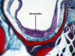
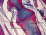
The eyeball is formed by the sclera
(stained green in the right picture), which forms a relatively thick ring in
front of the iris. Externally the sclera is covered by the thin conjunctiva
(stained purple in the right picture). The transparent part of the sclera in
front of the pupil is called the cornea. In the picture farthest to the
right you can see the conjunctival sac. This fold allows for the movement of
the eyes, preventing the penetration of dust and other objects between the sclera
and the eye-socket.
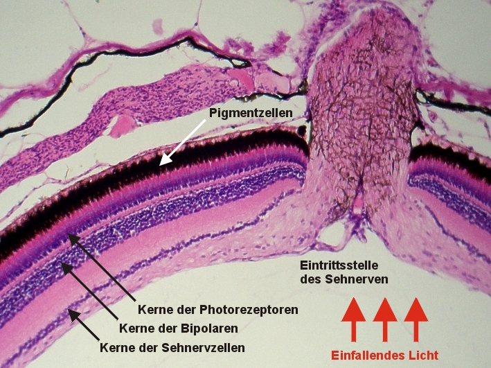
The retina of vertebrates
is a protruded part of the brain - the optic nerve should therefore be properly
named "optic tract", as it connects two parts of the brain.
 |
 |
 |
 |
|
|
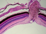
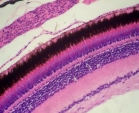
Retina
HE-staining |
Retina
HE-staining |
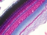
Retina
AZAN-staining |
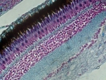
Retina
MASSON-GOLDNER |
|
 |
 |
|||
| Retina Scheme |
Loligo, everse
Retina HE-staining |
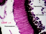
The retina of vertebrates is an "inverted retina" because the
photoreceptors are facing away from the light: the light must first pass all
layers of the retina before it falls on the photoreceptors. This arrangement
is a consequence of embryonic eye development, from the optical point of view
it is a disadvantage. For comparison the retina of a squid (Loligo)
is shown - here the photoreceptors are oriented towards the light, it is an
"everse retina".
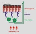
Even within the retina there is
a substantial preprocessing of images: edges are highlighted, in low-light receptors
are increasingly interconnected to form "clusters" (lower resolution,
increased sensitivity to light). This preprocessing is mainly done by the bipolar
cells (compare scheme), but also by the so-called horizontal cells
and the amacrines (not shown in the scheme). As the number of cells of
the optic nerve is much smaller than the number of photoreceptors, the resolution
of the image sent to the brain is much lower than expected - this restriction
of information is important for a quick analysis of images.
Behind the retina is the two-layered
pigment epithelium, like the retina part of the brain. The pigment melanin
prevents unwanted light reflections, but the biochemical cooperation between
the photoreceptors (rods and cones) and the adjacent cells of the pigment epithelium
is of more importance: During the process of light perception the receptors
constantly give off tiny bubbles, which are absorbed by the pigment cells. The
released substances are biochemically altered and the receptors constantly re-supplied.
If the retina loses contact to the pigment epithelium (retinal detachment),
this results in immediate blindness in the affected area. If this contact is
restored through surgery, blindness disappears within a few hours.
At the entrance of the optic nerve
receptors are missing - this is the "blind spot". But even
with monocular vision we do not notice this blind spot. This is so because the
picture we imagine to see only exists in our brain - the eye only provides the
"raw material". In fact, the optical quality of the eye is rather
poor, only a few degrees of a visual angle can be seen clearly.
A special feature of the vertebrate eye is the "fovea centralis",
a part of the retina a few millimeters in diameter, where there do not exist
any bipolar cells and thus a one-to-one connection between the photoreceptors
(usually cones) and the optic nerve cells is achieved (the place of sharpest
vision). In fish this fovea is missing, and light-dark-sensitive rods are predominant,
since fish are much more sensitive to light.
 |
 |
 |
| Lateral line
AZAN staining |
Lateral line
HE staining |
Taste bud MASSON-GOLDNER |
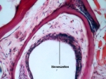
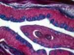
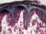
In the haed of fish there exists
a channel system with openings on the outside, which will continue from there
along the flanks, the "lateral organ". At the bottom of these
channels therev are groups of mechanoreceptors, which enable fish to
perceive water currents.
There are also "taste buds"
in the area of the head. They help fish to detect food, but they also have a
warning function. In land animals such receptor groups are restricted to the
oral cavity, usually confined to the tongue. In fish you see them also on the
surface of the body!
 |
 |
 |
 |
| Mucous
cells AZAN staining |
Bone
development AZAN staining |
Muscles
AZAN staining |
Intestinal
wall MASSON-GOLDNER |
The skin of the fish is covered
with mucus which is produced by special mucilage cells. Particularly
large cells of this type are found in the mouth, mucus becomes stained blue
when using the AZAN staining.
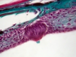
In small fish bone development is usually incomplete. The picture demonstrates how bones develop: First the shape of the bone is moulded by cartilage (stained blue), then the cartilage is replaced by bone (stained red). Real bone structures with typical HAVERSIAN SYSTEMS are absent here, as well as massive depositions of calcium carbonate and calcium phosphate (exact: hydroxyapatite).
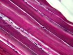
The next picture shows the typical
skeletal muscles of vertebrates. The muscle cells are polyenergid, they
have numerous mural cell nuclei. Inside the muscle cell are bundles of contractile
elements (fibrils). Each fibril shows a striated pattern. Since the fibrils
are arranged in a parallel structure, similar structural elements are on the
same level and so the cell shows striation.
Finally the upper series of pictures shows the bowel wall which contains a significant proportion of connective tissue (stained blue-green). Embedded in this tissue are smooth muscle cells (stained purple).Superposed is the mono-layered inner epithelium, which consists of elongated cells (stained mauve).
 |
 |
 |
 |
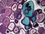
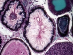
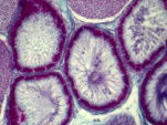
| Testicle MASSON-GOLDNER |
Testicle MASSON-GOLDNER |
Testicle AZAN-staining |
Testicle AZAN-staining |
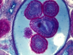
The primary sexual organs are particularly interesting. The testicle consists of numerous channels, where sperm cells mature. In viviparous fish, such as the guppies, sperm cells are glued together to spermatophores, which are then transferred to the female (internal fertilization). Spermatophores are shown in the image on the far left (surrounded by green-colored mucus) and the image on the far right.
Copyright: webmaster@mikrohamburg.de